Different Sized Pupils (Anisocoria) – at a glance
|
What is anisocoria?
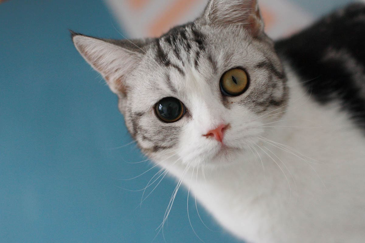
The pupils are the black opening located in the center of the eye, which expands and contracts depending on the amount of light. In low light, they expand, to let more light into the eye, in bright light, they contract to let less light in. Normally the pupils expand or contract together, with both either being dilated (large) or constricted (small).
Pupils of different sizes are medically known as anisocoria (ann-eye-so-CORE-ee-uh). The condition occurs for several reasons, and it may or may not be accompanied by other symptoms, depending on the underlying cause.
Causes
Anisocoria can be neurological or ocular and causes can range from harmless to life-threatening.
- Glaucoma: Increase in pressure inside the eye.
- Anterior uveitis: Inflammation of the uvea, the pigmented layer of the eye.
- Corneal ulcers: Damage to the surface layer of the cornea.
- Spastic pupil syndrome: Infection with the feline leukemia virus can cause this condition, and it may alternate between both eyes.
- Oculomotor nerve paralysis: The oculomotor nerve (cranial nerve III) is responsible for eye movement including constriction of the pupil; damage can occur due to systemic diseases such as diabetes or hypertension or due to head injury, tumours or aneurysms.
- Retinal detachment: A severe and sight-threatening disorder that occurs when the retina (the thin layer of tissue at the back of the eye) lifts or pulls away from the retinal pigment epithelium which provides nourishment and oxygen.
- Tumours: Brain, eye or optic nerve tumours.
- Horner’s syndrome: A condition causing drooping of the upper eyelid and constriction of the pupil caused by paralysis of the cervical sympathetic nerve supply.
- Stroke: A rupture or blockage of a blood vessel in the brain resulting in a loss of blood supply to an area of the brain affecting control of the ocular nerves.
- Head trauma: Can cause bleeding inside the brain which results in increased pressure in the skull.
- Iris atrophy: The iris is the coloured part of the eye and thinning of these cells can lead to a change in pupil size in the affected eye. This occurs most often in senior cats.
- Certain medications: Such as atropine which dilate the pupils. If the ocular medication has been used on one eye, only that pupil will dilate, however, systemic medications which can also cause pupil dilation will work on both of the pupils.
Symptoms
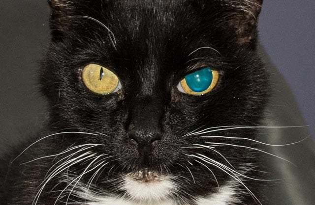
The primary symptom is uneven pupils, other symptoms secondary to the cause of anisocoria may include:
- Pain in the eye
- Change in the color of the eye, redness, or cloudiness
- Change to the position of the eye in the socket
- Abnormal eye movement
- Drooping eyelid
- Head tilting
- Loss of vision
- Confusion
- Retraction of the eye within the socket
Diagnosis
Your veterinarian will perform a physical examination of your cat and obtain a medical history from you including any medications or drops your cat is on if he has recently experienced trauma (such as a fall, hit by a car etc.) and other symptoms you may have noticed.
Physical examination:
A complete physical checkup and detailed examination of the eyes to look for signs of trauma including ulcers and scratches, abnormal pupil shape (dyscoria), and evaluate for obvious signs of disease.
He will need to determine if the affected pupil is abnormally dilated (mydriasis) or abnormally constricted (miosis). The room will be darkened to see if the constricted (smaller) pupil dilates (increases), and shining a bright light into the eyes to see if the pupil(s) constrict, as they should do.
Secondly, he will need to determine if the cause is an eye disorder or neurological.
Diagnostic workup:
- Baseline tests: Biochemical profile, complete blood count, and urinalysis to evaluate the overall health of your cat. These tests usually come back normal.
- Tonometry: This test measures the pressure in the eyes using a tonometer to check for glaucoma.
- Ultrasound: – Eye ultrasound to look for tumours or lesions in the eyes.
- Fluorescein staining: This involves placing a few drops of fluorescein in the eye to look for corneal ulcers.
- CT scan: An advanced non-invasive medical imaging technique that uses a combination of x-ray imaging and a digital computer to produce detailed 3-dimensional to look for lesions or tumours in the brain.
- Electroretinography (ERG): A measurement of the electrical responses to a light stimulus of the retinal photoreceptors (rods and cones), inner retinal cells and ganglion cells to evaluate the function of the retina at the back of the eye. The pupils are dilated with the use of medications and electrodes are placed on the cornea, bulbar conjunctiva or the skin below the lower eyelid. Flashes of light entering the eye trigger small amounts of electricity in the cells of the retina which are measured to determine how well they are working.
- Schirmer’s test: A paper test strip is placed between the palpebral conjunctiva of the lower eyelid and the bulbar conjunctiva of the eye to measure the production of tears.
- Neurological examination: The neurological examination evaluates the cat’s mental status, cranial nerves, motor examination, sensory examination and coordination.
- ELISA (enzyme-linked immunosorbent assay) or IFA (immunofluorescence assay): These blood tests detects antigen to the feline leukemia virus.
If your veterinarian can’t determine the cause, he will refer your cat to a veterinary ophthalmologist.
Treatment
The treatment of anisocoria will be tailored to address the underlying cause. Withdrawal of medications affecting the pupils. Antibiotics will be prescribed for bacterial infections. Where possible, surgery for tumors, and/or radiotherapy or chemotherapy as a follow-up. There are a number of medications that can be prescribed to manage glaucoma by reducing aqueous humour production or by increasing the outflow of intraocular fluid from the eye.
Secondary therapies:
There is no cure for feline leukemia; however, supportive care can increase the cat’s lifespan. This may include keeping your cat indoors, stress-free, changing his vaccine schedule, antibiotics to manage secondary infections, vitamins, antiviral drugs. ACE inhibitors, beta-blockers and calcium channel blockers to control high blood pressure as well as a low sodium diet. Horner’s syndrome usually resolves itself, in some cases, the veterinarian may treat symptoms with medications to dilate the pupil.
Prognosis
The prognosis varies depending on the cause. Seek veterinary attention immediately if you notice your cat has different-sized pupils.
Frequently Asked Questions
Why are my cat’s pupils different sizes?
- There are various causes of anisocoria including:
- Corneal ulcers
- Intra-cranial Disease
- Glaucoma
- Uveitis
- A disease of the Retina
- Iris atrophy
- Iris defects that are congenital
- Cancer
- Spastic pupil syndrome
- Infectious Disease
- Pain
What was the cause for such a dramatic difference in pupil size?
- Pain, inflammation, or damage affecting the nervous system input to the eye is the cause of the change in pupillary size.
How can I check my cat for glaucoma?
- Your veterinarian will use a tonometry device to assess your cat’s intraocular pressure.
Can anisocoria in cats be fatal?
- Anisocoria itself is a manifestation of an underlying disease process. Many are not fatal but infectious diseases or cancers may be.
Is anisocoria in cats painful?
- Anisocoria itself is not painful but the underlying problem causing anisocoria may be. Diseases like corneal ulcers, glaucoma, ocular trauma, and uveitis can be very painful for cats.
Can anisocoria in cats go away on its own?
- Sometimes anisocoria can improve without intervention. This is true if it is secondary to minor trauma or discomfort. Otherwise, it is unlikely to improve unless the underlying cause is treated.
When should I worry about unequal pupil sizes in cats?
- While anisocoria is rarely a life-threatening emergency (unless caused by ocular or head trauma) it can indicate something more serious is happening to your cat. Diseases like uveitis, glaucoma, and corneal ulcers can put your cat’s ocular health and vision at risk and require prompt intervention. Anytime anisocoria is a new finding, veterinary care should be sought.
Authors
-
by
-

-