What is pneumothorax?
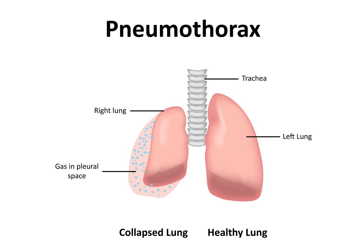
Pneumothorax (new-mo-thorax) is an abnormal accumulation of air in the pleural cavity which is between the lungs and the chest wall. Usually, there is a small amount of serous fluid in the pleural space which acts as a lubricant during breathing. Air entering the pleural space means there is less room for the lungs to expand when the cat inhales and causes the lung(s) to collapse (which is why the term collapsed lung is often used when describing pneumothorax).
It may be primary or secondary. Primary occurs in otherwise healthy cats; secondary is usually the result of an underlying condition.
Pneumothorax may develop in cats of any age, sex or breed. Cats who roam (particularly intact males) may be at higher risk of traumatic pneumothorax due to their susceptibility to roam and get into fights.
Causes
There are three classifications of pneumothorax in cats depending on the cause.
- Traumatic
- Spontaneous
- Iatrogenic
Pneumothorax can affect one or both of the lungs, bilateral (both lungs) is more common and can be open or closed. Air enters the pleural space in one of two ways, from a penetrating wound through the thoracic wall, which allows air to enter from the outside (open), or air leaking from the respiratory tract or esophagus into the pleural space (closed).
Open pneumothorax occurs when there is an unsealed opening in the chest wall and is most commonly associated with traumatic pneumothorax. Air can enter the pleural space from the external environment during inhalation. This results in a loss of negative pressure in the pleural cavity which is necessary to keep the lung expanded; as a result, the lung collapses. Air is pulled into the chest cavity through the opening when the cat inhales.
A rare complication can occur in some cats where a one-way valve develops so when the cat inhales air is sucked in but is unable to exit when the cat exhales, this is known as tension pneumothorax and is life-threatening. It can occur with both traumatic and spontaneous pneumothorax.
Traumatic pneumothorax:
This is the most common cause of pneumothorax in cats, common causes include:
- Penetrating wounds from gunshots, bites, stab wounds which allow air from outside into the pleural cavity
- Fractured ribs can penetrate the lungs
- Trauma to the lungs allows air to escape into the surrounding area such as a broken rib
- Trauma from a vehicle
- Fall from a height
- Lung infection
Spontaneous pneumothorax:
This type of pneumothorax develops without trauma and is always closed and can be classified as primary or secondary. Primary occurs when there is no apparent underlying cause.
- Pulmonary bleb rupture or bullae emphysema
- Cancer
- Heartworm
- Lungworm
- Lung flukes
- Pulmonary abscess
- Pneumonia
- Lung lesions due to cysts, abscess, parasites
- Grass awn migration
- Asthma
- Rupture of the bronchus, trachea, lung or esophagus
Iatrogenic pneumothorax:
Medically induced pneumothorax occurs as a result of surgical procedures such as:
- Bronchoscopy
- Thoracocentesis
- Thoracostomy tube
- Intubation
- Lung surgery
Symptoms
Difficulty breathing is the most common sign although the extent of symptoms may vary from subtle to severe depending on the severity of the disease. Generally, traumatic pneumothorax will be obvious. However, this may not be the case with spontaneous pneumothorax, particularly if the leak is slow.
- Cyanosis (blue-tinged gums)
- Dyspnea (difficulty breathing)
- Open-mouthed breathing/panting
- Exercise intolerance
- Tachypnea (rapid, shallow breathing)
- Restlessness
- When lying down your cat may lie flat (sternal recumbency)
- Anorexia (loss of appetite)
Diagnosis
The veterinarian will perform a complete physical examination of the cat which will include listening to the chest for abnormal, harsh or decreased lung sounds.
Diagnostics:
- Baseline tests: Biochemical profile, complete blood count, and urinalysis usually return normal results but can be of use in evaluating organ function of cats who have been involved in a trauma such as a road accident or fall.
- Thoracocentesis: A procedure that is both diagnostic and therapeutic. The veterinarian inserts a needle through the chest wall and into the pleural space and removes the air. This both shows that pneumothorax is present as well as provides relief from symptoms.
- Chest x-ray: Confirmation of pneumothorax is made via chest x-rays which can also provide additional information such as the extent of pneumothorax as well as possible causes.
- CT scan: An advanced non-invasive medical imaging technique that uses a combination of x-ray imaging and a digital computer to produce detailed 3-dimensional images to look for the bullae, blebs and lung lesions as it is more sensitive than x-rays.
- Echocardiogram: A non-invasive test that uses high-frequency sound waves to capture live images of the heart to examine the anatomy and function of the heart and valves to look for heartworms.
- Antigen or antibody tests: To look for antigen or antibodies to heartworm or microfilaria.
- Fecal examination or tracheal aspiration: To look for lungworm or lung fluke eggs or larvae.
Treatment
The goal of treatment is to address the underlying cause and provide supportive care to relieve symptoms. Medical management may only be necessary in cases of a small and spontaneous pneumothorax. Small tears should heal on their own, and over time, the air will reabsorb into the bloodstream.
- Thoracocentesis (see above in diagnosis) to remove trapped air.
- Tube thoracostomy (chest tube) where recurrent thoracocentesis has occurred. A thin tube is inserted into the pleural space to remove air; the tube remains in place so that air can be removed intermittently or continually via a suction unit.
- Surgery will be necessary to treat cats with penetrating wounds, grass awns or tumours.
- Surgery to treat blebs, bullae and lung abscess if medical management isn’t successful.
- Oxygen therapy for cats whose oxygen levels are low. This can also help to push the free air in the pleural cavity into the pleural blood vessels.
- Analgesics to relieve pain.
Once the cat is stable, the veterinarian can focus on treating the underlying cause.
Home care
Administer medications (if prescribed) as directed by your veterinarian.
Confine the cat in a large cage or small room during recovery.
Careful monitoring of your cat is essential during this time. If you notice he is developing breathing difficulties again, he needs immediate veterinary attention.
Author
-
by
-