At a glanceAbout: Saddle thrombosis is a life-threatening condition that occurs when a blood clot breaks off from a larger clot in the heart and travels down the aorta, lodging at the junction of the iliac arteries of the hind legs. Causes: The most common cause is heart disease, typically hypertrophic cardiomyopathy. Symptoms:
Treatment: Pain relief, IV fluids to correct electrolyte derangements and thrombolytics to dissolve the clot. |
About
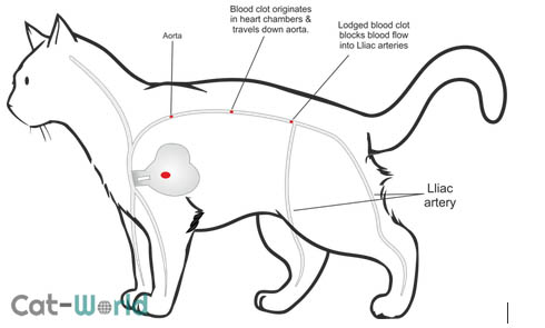 Saddle thrombosis (aortic thromboembolism) is a life-threatening condition in which a blood clot (thrombus) forms in the heart and then all or part of it dislodges and travels down the abdominal aorta where it lodges at the junction of the iliac arteries of the hind legs which blocking blood flow to the hind limb(s).
Less common locations the emboli may travel to include the arteries supplying the kidneys, lungs, gastrointestinal tract, forelegs, and brain. However, this article will focus on saddle thrombosis only which affects the rear legs, the term saddle thrombosis is due to the emboli lodging in the junction and draping down into the iliac veins, giving it the appearance of a saddle. Studies show between 69-90% of cats with thromboembolism have heart disease and up to 1/3rd of cats with heart disease will develop a blocked artery due to thromboembolism. Other causes include neoplasia and hyperthyroidism.
The aorta is the largest artery in the cat’s body supplying oxygenated body to the organs. It originates in the left ventricle of the heart, travelling down the abdomen before splitting into the left and right iliac arteries as well as the caudal vein which supplies blood to the back legs and tail. The junction where the aorta splits is known as the aortic bifurcation. A thrombus is a blood clot made up of platelets and fibrin, and an embolus is something that travels through a blood vessel until the vessel is too small to let it continue.
Causes
Saddle thrombosis is devastating, as there is no warning. The majority of cases are due to an underlying heart condition (usually hypertrophic cardiomyopathy) which in many cases many cat owners and veterinarians are unaware of until aortic thromboembolism occurs. Hyperthyroidism and neoplasia may also cause aortic thromboembolism.
Almost all blood clots form in the left atrium of the heart which may be due to a thickening of the atrium wall can cause blood to pool, leading to the formation of these clots, or increased turbulence. At any time a clot formed in the heart can dislodge and travel along the aorta before becoming wedged in the narrower iliac arteries.
Once blocked, blood can no longer pass into the iliac arteries, which means tissues in the legs are no longer receiving am adequate (or any) oxygen (ischemia), a cascade of biochemical reactions within the cells follow, leading to toxic by-products which ultimately cause cell death. Unfortunately, it doesn’t end there, once the blockage clears, all the toxic by-products which have built up are released back into the circulatory system (reperfusion injury).
Saddle thrombosis can develop in cats of any age or breed; one study found the mean age was 12 years.
Symptoms
Clinical signs will depend on the location of the embolism. Saddle thrombosis is typically identified by the 5 P’s:
- Pain: Affected cats are in extreme pain with distressed howling/vocalisation and anxiety common symptoms
- Paralysis or paresis (weakness): The most common location of the embolism is the terminal aorta, most cats present with complete paralysis or weakness/partial loss of the hind limbs
- Pulselessness: There may be a faint pulse in some cats if there isn’t a complete blockage).
- Poikilothermic: The limbs are cold to the touch.
- Pallor
Other findings can include:
- Vomiting (before ATE)
- Gait abnormalities
- Restlessness initially, presumably due to pins and needles type feelings in the leg(s) as blood supply diminishes.
- Tachypnea (rapid/shallow breathing) with open-mouthed breathing due to pain or heart disease.
- The paw pads may appear cyanotic (blue).
- Hard and painful muscles.
Aortic thromboembolism is a medical emergency and requires immediate veterinary attention.
Diagnosis
Your veterinarian will perform a complete physical examination of your cat and obtain a medical history from you. During the exam, the veterinarian will check for a pulse in the rear limbs which may be faint or may not be there at all. The leg(s) and feet will feel cold to the touch with a blue-tinged skin tone on the paw pads. If clipped beyond the quick, claws on the affected limb fail to bleed.
Diagnostic workup:
- Baseline tests: Complete blood count and biochemical profile which may reveal hyperglycemia (increased blood glucose levels) due to stress, elevated muscle enzymes creatine kinase.
- Stethoscope examination: Abnormal cardiac sounds are a common finding when the veterinarian listens to the heart with a stethoscope (auscultation) due to underlying heart conditions.
- Peripheral blood glucose test: The veterinarian will obtain a blood sample from the affected limbs to check the glucose level, which should be reduced in the limbs of cats with aortic thromboembolism compared to normal values from the biochemical profile.
- High blood calcium and metabolic acidosis if reperfusion has occurred.
- Doppler ultrasound: A noninvastive test using ultrasound waves to estimate blood flow through the blood vessels by bouncing ultrasound (high frequency sound waves) off red blood cells.
- Chest x-rays and/or echocardiogram (ultrasound): To evaluate the size and shape of the heart.
- Doppler ultrasound: A noninvasive test that can be used to estimate the blood flow through your blood vessels by bouncing high-frequency sound waves (ultrasound) off circulating red blood cells. A regular ultrasound uses sound waves to produce images, but can’t show blood flow.
Treatment
Immediate:
- Stabilise cats with congestive heart failure which may include oxygen therapy and Furosemide (a diuretic) to remove fluid build-up.
- Fluid therapy can help to counteract the toxic effects of byproducts produced during ischemia. Care must be taken when giving fluids to a cat with heart disease.
- Analgesics such as hydromorphone or buprenorphine to relieve pain.
Management:
- Medications to dissolve the blood clot (thrombolytics).
- Anticoagulant therapy such as heparin or aspirin to thin the blood and help stop additional blood clots from forming.
- Glucose, insulin and in some cases, sodium bicarbonate to treat hyperkalemia.
- The cat will require cage rest and around the clock care during recovery.
It is important to treat the underlying heart condition once your cat has recovered from saddle thrombosis.
Treatment is often unsuccessful. Blood thinners and thrombolytics run the risk of causing bleeding complications. Even if the cat recovers from the initial event, the risk of another clot forming is extremely high. On top of that is usually an underlying heart condition. Many owners elect to euthanise cats with aortic thromboembolism due to the poor outcome.
Author
-
by
-